Damn you cats: Squirrels ‘dropping dead from trees’ from Toxo outbreak

Toxoplasma gondii, a zoonotic protozoan parasite for which felids are the only definitive hosts, can infect humans and other warm-blooded animals.

cats.sink.jun.13Transmission usually occurs orally from oocysts shed by felids in water and on food, through tissue cysts in undercooked meat, or transplacentally. In particular, young cats shed oocysts that can sporulate and become infectious within a day, depending on temperature and humidity. Sporulated oocysts can survive in moist soil for months to years (1).

In September 2014, the number of dead squirrels reported to the Dutch Wildlife Health Centre and the Dutch Mammal Society increased suddenly. The red squirrel (Sciurus vulgaris) is the only species of squirrel endemic to the Netherlands. Members of the public claimed that squirrels were “dropping dead from trees.” Subsequently, the public was encouraged to report and submit dead squirrels. A total of 187 animals were reported through October 2014, of which 37 were submitted for necropsy. Necropsy included macroscopic examination; cytologic analysis of liver, spleen, lungs, and intestinal contents stained with hemacolor (Merck, Darmstadt, Germany); and histologic examination of samples of various organs fixed in formalin, embedded in paraffin, cut into 4-μm sections, and stained with hematoxylin and eosin.

For 8 adult animals, body condition (based on degree of fat storage and muscle development) was good; 12 juveniles were in poor condition. Typically, the trachea contained foam, and lungs were hyperemic and edematous. The liver was enlarged and pale, and the spleen was enlarged. In 13 animals, numerous small crescent-shaped organisms, with eccentrically placed nuclei consistent with tachyzoites of T. gondii, were identified by cytology in lung, liver, and spleen (2). Main histopathologic findings were pulmonary interstitial lymphoplasmocytic and neutrophilic infiltrates with edema and numerous intra-alveolar macrophages (17/20) and multifocal lymphoplasmocytic infiltrates with necrosis in the liver (13/20). Extensive splenic necrosis was occasionally observed (4/20). Intestines contained mild plasmacytic infiltrates. Numerous tachyzoites consistent with T. gondii were present in alveolar macrophages and epithelial cells, splenic macrophages, and hepatocytes. Duplicate slides were stained immunohistochemically by using polyclonal antibodies against T. gondii following a standard ABC protocol (3). Organisms stained for T. gondii in liver, spleen, lungs, and intestine. Toxoplasma was not detected in any brain. DNA was isolated (DNeasy Blood and Tissue Kit; QIAGEN, Hilden, Germany) from tissues of 14 squirrels and tested by quantitative PCR (1); T. gondii DNA was detected in 13. We successfully sequenced the T. gondii GRA6 gene for 11 squirrels and identified sequences to clonal type II T. gondii previously identified in sheep from the Netherlands (GenBank accession no. GU325790) (4). Incidental findings in the animals tested were encephalitis (2/20), coccidiosis (5/20), trauma (6/20), myocarditis (4/20), nephritis (1/20), lymphadenitis (1/20), and intestinal (3/20) and external (5/20) parasites.

The remaining 17 animals showed >1 of the following pathologic conditions: hemorrhages consistent with trauma (12/17), mild to severe intestinal coccidiosis (12/17), pneumonia (3/17), splenitis (1/17), Taenia martis cysticerci (1/17), and external parasites (8/17). Immunohistochemistry results for all 17 were negative for T. gondii.

grey.squirrel.eatOn the basis of necropsy and molecular findings, we conclude that 20 of 37 examined squirrels died of disseminated T. gondii type II infection. These animals included adults and juveniles and were not restricted to specific geographic areas. The remaining animals died of trauma (12/17) or other causes (5/17).

Red squirrels are susceptible to T. gondii, and infection can lead to death. However, in our sample, the proportion of squirrels that died of toxoplasmosis (>50%) was higher than in other studies (≈16%) (5,6,7). The apparent increase in squirrel deaths and unexpectedly high proportion of fatal T. gondii infections suggests a toxoplasmosis outbreak among red squirrels. Possible explanations for this surge in cases include increased exposure to the parasite, increased susceptibility to infection, or increased virulence of the pathogen. Clonal T. gondii type II, the strain most frequently involved in human cases and endemic to Europe and North America, was identified. An increased virulence of the pathogen could not be proven (8). On the basis of lymphoid hyperplasia in the spleen and lymph nodes, affected squirrels had no signs of immunosuppression. Thus, the most likely explanation is increased exposure to the parasite.

Sources of infection for red squirrels are not known; however, oocysts shed in cat feces may contaminate the nuts, fungi, shoots, and berries that constitute the diet of the squirrel. Stray, unspayed cats are common in the Dutch countryside. More than 3 million domestic cats (Felis domesticus) exist in the Netherlands, including several tens of thousands of free-roaming cats that reproduce (9). Determining the exact source of infection is important because humans also harvest wild fruits, nuts, and fungi from these areas. This outbreak highlights that contamination of the environment with T. gondii oocysts is of concern not only from a public health viewpoint but from a biodiversity perspective as well (1,10).

Marja Kik Comments to Author , Jooske IJzer, Marieke Opsteegh, Margriet Montizaan, Vilmar Dijkstra, Jolianne Rijks, Andrea Gröne, and Jooske IJzer

Author affiliations: Utrecht University, Utrecht, the Netherlands (M. Kik, J. IJzer, M. Montizaan, J. Rijks, A. Gröne); Dutch National Institute for Public Health and the Environment, Bilthoven, the Netherlands (M. Opsteegh); Dutch Mammal Society, Nijmegen, the Netherlands (V. Dijkstra)


Toxoplasma gondii in Wild Red Squirrels, the Netherlands, 2014

Emerging Infections Diseases, Volume 21, Number 12, December 2015

http://wwwnc.cdc.gov/eid/article/21/12/14-1711_article

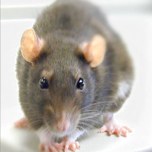
Why inspectors fret about rats: Rodents as hosts of infectious diseases

Rodents are recognized as hosts of more than 60 zoonotic diseases that represent a serious threat to human health (Meerburg et al. 2009, Luis et al. 2013). This special issue emerges from a workshop organized in Bangkok at the Faculty of Veterinary Medicine of Kasetsart University and supported by the French ANR project CERoPath (Community Ecology of Rodents and their Pathogens in a Southeast Asian changing environment), which aimed at better understanding the relationships between rodent-borne diseases, rodents and their habitats using intensive field works, serology, and molecular screenings.

sq-willard-crispin-glover-rat-nlThe main objective of this workshop was to join ecologists, biologists, and epidemiologists to give an overview on the importance of rodents as hosts and reservoirs of parasitic and infectious diseases. Most of presentations given in the workshop focused in Southeast Asia, a hotspot of both infectious emerging diseases (Coker et al. 2011) and biodiversity at threat due to dramatic changes in land use (Morand et al. 2014).

A first challenge is related to the invasion or range expansion of rodents. The black rat (Rattus rattus), Norway rat (Rattus norvegicus), Asian house rat, (Rattus tanezumi) and Pacific rat (Rattus exulans) like the house mice (Mus musculus), have dramatically expanded their geographic range as a consequence of human activities (Aplin et al. 2011). All of these Rattus species originated in Asia, and can be found in sympatry due to their synanthropic behavior (McFarlane et al. 2012). These rodents have been implicated (Kosoy et al., this issue), and still are implicated (Kuo et al. 2011), in the emergence and spread of plague, murine typhus, scrub typhus, leptospirosis, hantavirus hemorrhagic fever, among others. A better comprehension of the range extension mechanisms and consequences in term of infectious diseases’ risks would require investigation of the genetics and immunology of these rodent species (Himsworth et al., this issue) as well on the ecological interactions among pathogens, vectors, and rodents. Gutiérrez et al. (this issue) in their review attempt to summarize and bridge some knowledge gaps in the transmission and distribution routes, and in the dynamics and composition of Bartonella-infection in rodents and their flea parasites.

A second challenge is that in order to better understand disease ecology and parasite transmission, it must be considered that not all hosts are equally involved in parasite transmission as some species (and individuals) can be responsible for a disproportionate number of transmission events (Paull et al. 2012). Some habitats or landscapes may also disproportionally enhance transmission or persistence of parasite and/or vector (Bordes et al. 2013). Indeed, by carrying and disseminating parasites across multiple habitats, generalist or synanthropic rodents could enhance both host-switching and spill-over to other rodent reservoirs and directly or indirectly to humans.

rat.restaurantSeveral articles in this special issue suggest high diversity of pathogens and parasites circulating among diversified communities of rodents across various habitats, such as Rickettsia spp. in Taiwan (Kuo et al., this issue), Bartonella spp. in Vietnam (Hoang et al., this issue) and in Thailand (Jiyipong et al., in this issue), leptospirosis in Vietnam (Hoang et al., this issue), zoonotic viruses in Vietnam (Cuong et al., this issue), and zoonotic helminths in Thailand (Chaisiri et al., this issue).

This special issue calls for a more thorough investigation of rodent-borne diseases taking into account the ecology of rodents in their habitats. Taken together, the studies presented in this special issue stress the need to identify the mechanisms affecting pathogen diversity and infection within and between rodent species and to explain why some rodent species seem to be more resilient to greater habitat disturbance. Future research should monitor a variety of rodent pathogens simultaneously, including directly-transmitted specific pathogens and vector-borne pathogens with varying degrees of specificity.

Vector-Borne and Zoonotic Diseases. January 2015, 15(1): 1-2.

Morand S., Jittapalapong S., and Kosoy Michael

http://online.liebertpub.com/doi/abs/10.1089/vbz.2015.15.1.intro#utm_source=ETOC&utm_medium=email&utm_campaign=vbz

Tourists in India face threat of zoonotic diseases

Tourists are endangering their health and safety by interacting with elephants that are not screened for any zoonotic diseases, with no vaccination and treatment records with the mahouts, observed PETA.

600full-the-elephant-man-screenshotA team of four veterinarians assessed the health of elephants and various issues came to light during the inspection.

The overall assessment of 34 elephants at Amber was carried out with the International Union for Conservation of Nature (IUCN) checklist, ‘Asian Elephant Body Condition Index’, ‘Elephant Physical Examination Record’ and ‘Elephant Medical History’.

Many serious epidemic diseases are zoonoses that originated in animals. These include rabies, Ebola virus and influenza. In a systematic review of 1,415 pathogens known to infect humans, 61% were zoonotic.

“The owners/mahouts did not have any record of vaccination and treatments carried out in the past for their elephants. They did not maintain vaccination register nor the disease and treatment register,” said Dr Manilal Valliyate from PETA. 

Whole genome sequencing reveals potential spread of Clostridium difficile between humans and farm animals in the Netherlands, 2002 to 2011

Farm animals are a potential reservoir for human Clostridium difficile infection (CDI), particularly PCR ribotype 078 which is frequently found in animals and humans. Here, whole genome single-nucleotide polymorphism (SNP) analysis was used to study the evolutionary relatedness of C. difficile 078 isolated from humans and animals on Dutch pig farms.

UQ.piggeryAll sequenced genomes were surveyed for potential antimicrobial resistance determinants and linked to an antimicrobial resistance phenotype. We sequenced the whole genome of 65 C. difficile 078 isolates collected between 2002 and 2011 from pigs (n = 19), asymptomatic farmers (n = 15) and hospitalised patients (n = 31) in the Netherlands. The collection included 12 pairs of human and pig isolates from 2011 collected at 12 different pig farms. A mutation rate of 1.1 SNPs per genome per year was determined for C. difficile 078. Importantly, we demonstrate that farmers and pigs were colonised with identical (no SNP differences) and nearly identical (less than two SNP differences) C. difficile clones.

Identical tetracycline and streptomycin resistance determinants were present in human and animal C. difficile 078 isolates. Our observation that farmers and pigs share identical C. difficile strains suggests transmission between these populations, although we cannot exclude the possibility of transmission from a common environmental source.

Euro Surveill. 2014;19(45):pii=20954

Knetsch CW, Connor TR, Mutreja A, van Dorp SM, Sanders IM, Browne HP, Harris D, Lipman L, Keessen EC, Corver J, Kuijper EJ, Lawley TD

http://www.eurosurveillance.org/ViewArticle.aspx?ArticleId=20954

Reverse zoonotic disease transmission (zooanthroponosis): A systematic review of seldom-documented human biological threats to animals

Thanks to friend of the blog, Michael Batz, for flagging this article.

Background

sadie.sorenne.car.10Research regarding zoonotic diseases often focuses on infectious diseases animals have given to humans. However, an increasing number of reports indicate that humans are transmitting pathogens to animals. Recent examples include methicillin-resistant Staphylococcus aureus, influenza A virus, Cryptosporidium parvum, and Ascaris lumbricoides. The aim of this review was to provide an overview of published literature regarding reverse zoonoses and highlight the need for future work in this area.

Methods

An initial broad literature review yielded 4763 titles, of which 4704 were excluded as not meeting inclusion criteria. After careful screening, 56 articles (from 56 countries over three decades) with documented human-to-animal disease transmission were included in this report.

Findings

In these publications, 21 (38%) pathogens studied were bacterial, 16 (29%) were viral, 12 (21%) were parasitic, and 7 (13%) were fungal, other, or involved multiple pathogens. Effected animals included wildlife (n = 28, 50%), livestock (n = 24, 43%), companion animals (n = 13, 23%), and various other animals or animals not explicitly mentioned (n = 2, 4%). Published reports of reverse zoonoses transmission occurred in every continent except Antarctica therefore indicating a worldwide disease threat.

Interpretation

As we see a global increase in industrial animal production, the rapid movement of humans and animals, and the habitats of humans and wild animals intertwining with great complexity, the future promises more opportunities for humans to cause reverse zoonoses. Scientific research must be conducted in this area to provide a richer understanding of emerging and reemerging disease threats. As a result, multidisciplinary approaches such as One Health will be needed to mitigate these problems.

PLoS ONE 9(2): e89055

Messenger AM, Barnes AN, Gray GC

http://www.plosone.org/article/info:doi/10.1371/journal.pone.0089055


Is that an African or European swallow? Can’t kill all the birds, but can manage risk

Zoonotic enteric pathogenic bacteria can live in the intestinal tract of birds and can be transmitted to food animals or humans via fecal contact. In the present study, cecal samples were collected from 376 migratory birds from species birdsfilm460often associated with cattle during the fall migration in the Central Flyway of the United States. Brown-headed cowbirds (n=309, Molothrus ater), common grackles (n=51, Quiscalus quiscula), and cattle egrets (n=12, Bubulcus ibis) contained foodborne pathogenic bacteria in their ceca. Salmonella enterica was isolated from 14.9% of all samples, and Escherichia coli O157:H7 from 3.7%. Salmonella serotypes isolated included the following: Muenster, Montevideo, and Typhimurium.

Our data suggest that migratory birds associated with cattle could be a vector for zoonotic enteric pathogenic bacteria to be disseminated across long distances.’

Isolation of Escherichia coli O157:H7 and Salmonella from migratory brown-headed cowbirds (Molothrus ater), common grackles (Quiscalus quiscula), and cattle egrets (Bubulcus ibis)

 Foodborne Pathogens and Disease. -Not available-, ahead of print

Callaway, Todd R., Edrington, Tom S., and Nisbet, David J.

http://online.liebertpub.com/doi/abs/10.1089/fpd.2014.1800

Sanitizers not enough says UK; wash hands after visiting the farm

The UK Public Health Agency (PHA) is reminding families about the importance of washing hands after visiting the farm.

petting.zoo.handwash.10All animals naturally carry a range of organisms, some of which can be transmitted to people. Some organisms which may be contracted on farms present a serious hazard and can potentially cause severe infection, particularly in young children.

To reduce the risk of illness, both adults and children should wash their hands thoroughly using soap and water after they have handled animals or touched surfaces at the farm and always before eating or drinking.

Antibacterial hand gels and wipes are not a substitute for washing hands with soap and water, as gels and wipes may be unable to remove contamination in the way that running water can. However, using such gels after hand washing with soap and water may reduce further the risk of picking up these infections.


Dr Philip Veal, Consultant in Health Protection at the PHA, said: “Farm animals often carry a range of organisms which can be passed to children and adults. These organisms can include serious infections such as E. coli O157 which is extremely contagious and easily passed from animals to children and then within the household.

“Hand washing with soap and water will reduce the risk of picking up these infections, which can be particularly harmful to young children. By being aware and by doing these simple things we can help to avoid illness and enjoy a fun day out.”

Best practices for planning events encouraging human-animal interactions

03.Apr.14

Zoonoses and Public Health

G. Erdozain , K. KuKanich , B. Chapman  and D. Powell

http://onlinelibrary.wiley.com/doi/10.1111/zph.12117/abstract?deniedAccess

Educational events encouraging human–animal interaction include the risk of

zoonotic disease transmission. It is estimated that 14% of all disease in the US caused by Campylobacter spp., Cryptosporidium spp., Shiga toxin-producing Escherichia coli (STEC) O157, non-O157 STECs, Listeria monocytogenes, nontyphoidal Salmonella enterica and Yersinia enterocolitica were attributable to animal contact. This article reviews best practices for organizing events where human–animal interactions are encouraged, with the objective of lowering the risk of zoonotic disease transmission.

Animals can make humans poop: a systematic review and meta-analysis

Domestic animal husbandry, a common practice globally, can lead to zoonotic transmission of enteric pathogens. However, this risk has received little attention to date. This systematic review and meta-analysis examines the evidence for an association between domestic exposure to food-producing animals and cases of human diarrhea and specific enteric infections.

family.guy.diarrheaWe performed a systematic review of available literature to examine domestic livestock and poultry as risk factors for diarrhea and applied pre-determined quality criteria. Where possible, we carried out meta-analysis of specific animal–pathogen pairs.

We found consistent evidence of a positive association between exposure to domestic food-producing animals and diarrheal illness across a range of animal exposures and enteric pathogens. Out of 29 studies included in the review, 20 (69.0%) reported a positive association between domestic animal exposure and diarrhea. Domestic exposure to poultry revealed a substantial association with human campylobacteriosis (OR 2.73, 95% CI 1.90–3.93).

Our results suggest that domestic poultry and livestock exposures are associated with diarrheal illness in humans. Failure to ascertain the microbial cause of disease may mask this effect. Exposure to domestic animals should be considered a risk factor for human diarrheal illness and additional studies may identify potential mitigation strategies to address this risk. 

Human diarrhea infections associated with domestic animal husbandry: a systematic review and meta-analysis

Trans R Soc Trop Med Hyg, March 2014

Laura D. Zambrano, Karen Levy, Neia P. Menezes and Matthew C. Freeman

Better zoonoses surveillance needed in Nepal

A team of scientists from Belgium, Nepal, New Zealand, Switzerland and the Netherlands report in a study titled ‘The Burden of Parasitic Zoonoses in Nepal: A Systematic Review’ that the country needs a better surveillance system to accurately estimate animal-borne parasitic disease or ‘parasitic zoonoses (PZs)’.

The recent study estimates the public health burden of PZs in Nepal at 24,000 healthy pork tapewormsyears lost annually.

Neurocysticercosis caused by pork tapeworms affect the nervous system, cystic echinococcosis traced through dog tapeworms and congenital toxoplasmosis in which mothers infected with the toxoplasma parasite give birth to infected children, are the three top PZs in Nepal, according to the study.

The study estimates that Nepal loses 14,268 healthy years annually due to neurocysticercosis, 9,255 years due to congenital toxoplasmosis, and 251 due to cystic echinococcosis. Numerous data sources were analysed in the study to examine the relevance and importance of such infections.

“Nepal is considered endemic for at least 10 PZs, and might be endemic for seven others. Most of these diseases probably only have a small public health impact. However, neurocysticercosis and congenital toxoplasmosis are likely to impose an important burden to public health,” said the report.

One Health pioneer, James H. Steele, dies at 100

The potential for a disease to pass from animals to people had been understood for millenniums, but it was not until the late 18th century, when Edward Jenner developed a vaccine for smallpox, that someone found a way to prevent it. The idea that government could take a systematic approach to fighting disease in animals to protect people did not take hold until the middle of the 20th century, when, according to a N.Y. Times obituary, Dr. James Steele led the way.

He helped establish mass vaccination and prevention programs in the United States for diseases like rabies and bovine brucellosis. After setting up federal programs, he helped start them at the state james.steelelevel. He visited dozens of countries to start veterinary public health programs and to help trace specific diseases, like Rift Valley fever in Nigeria, where he traveled in the 1970s. He constantly looked beyond his immediate field: in 1964, he published a paper titled “The Socioeconomic Responsibilities of Veterinary Medicine.”

He participated via Skype this summer at the annual conference of the American Veterinary Medical Association in Chicago during a lecture series, “The James Steele Challenge: A Better World Through One Health.” It focused on his lifelong passion: convincing people that economic prosperity was rooted in animal, human and environmental health. Scores of students cite him as their mentor.

In 1942, a year after Dr. Steele received his doctor of veterinary medicine degree from Michigan State University, he became one of the first veterinarians to receive a master’s degree in public health from Harvard. In 1945 he started the veterinary public health program at the United States Public Health Service in Washington. In 1947, he and the unit moved to Atlanta, to what is now called the Centers for Disease Control and Prevention.

Two years after that he went to work for the office of the surgeon general as chief veterinary officer. He became the nation’s first assistant surgeon general for veterinary affairs in 1968 and deputy assistant secretary for health and human services in 1970. The formal name for the types of diseases Dr. Steele dealt in, which pass from animals to humans, is zoonoses. While he had strong science and field experience — his interest in pursuing a veterinary degree increased after he helped investigate a brucellosis outbreak in a lab at Michigan State in the 1930s — his special talent was in finding practical ways to address disease on a large scale.

“He had to take all that science and translate it into a disease control program,” said Dr. Peter Cowen, who teaches epidemiology and public health in the college of veterinary medicine at North Carolina State University. “Jim took the science and protected public health.”

James Harlan Steele was born on April 3, 1913, in Chicago to James Hahn Steele and the former Lydia Norquist. He grew up, all the way to 6-foot-7, in Chicago, and stayed there into his 20s, selling insurance to help his family before he entered Michigan State.

In 1971, he became a professor at the University of Texas School of Public Health in Houston.

“The impact of veterinary research through the years has been startling,” he wrote in the 1960s. “It has opened vast areas of continents to animal husbandry, given a base to many industries and improved human nutrition beyond expectation. Probably in no other creative area has an investment returned so great a dividend for mankind.”

He retired in 1983. His survivors include his wife, Brigitte; his sons, Michael, James and David; and four grandchildren. His first wife, the former Aina Oberg, died in 1969.