As I’ve written before, I didn’t really get turned onto learning until a 4th year university virology course in 1984 (I half since seen the prof several times and thanked him profusely).
John Oliver and his writing staff have done everything I’ve tried to do, but he has better writers who are also paid better.
Given that phages are able to destroy bacteria, they are of particular interest to science. Basic researchers from the Leibniz-Forschungsinstitut für Molekulare Pharmakologie (FMP) in Berlin are especially interested in the tube used by phages to implant their DNA (or RNA – dp) into bacteria. In collaboration with colleagues from Forschungszentrum Jülich and Jena University Hospital, they have now revealed the 3D structure of this crucial phage component in atomic resolution. The key to success was combining two methods – solid-state NMR and cryo-electron microscopy. The study has just been published in the journal Nature Communications.
With growing antibiotic resistance, phages have increasingly become the focus of research. Phages are naturally occurring viruses with a very useful property: they implant their DNA into bacteria and proliferate there until the bacterial cell is ultimately destroyed. This is why they are also referred to as bacteriophages (bacteria eaters). This approach has already been shown to fight multidrug-resistant bacteria. Last year, the case of a girl from England hit the headlines, when she was cured from a serious antibiotic-resistant infection using engineered phages.
However, the widespread use of phage therapy is still a long way off. Many of the underlying principles that are key to advancing this therapy are not yet understood. For example, little was previously known about the appearance of the exact architecture of the tube used by phages to implant their DNA into bacteria. Now scientists from the Leibniz-Forschungsinstitut für Molekulare Pharmakologie (FMP) in Berlin, together with colleagues from Forschungszentrum Jülich and Jena University Hospital, have managed to reveal the 3D structure of this crucial phage component in atomic resolution.
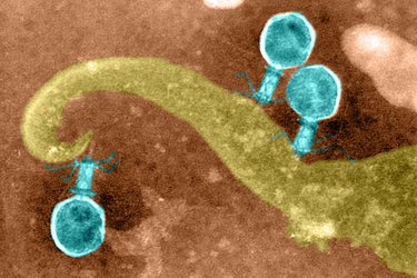
“The structure and flexibility of the DNA tube attached to the icosahedron-shaped capsid is somewhat reminiscent of a spinal column,” stated FMP’s Professor Adam Lange, describing one of the new findings. “It seems to be perfectly designed for transporting DNA.”
The researchers were able to gain insights into the structure and function of this sophisticated DNA transport pathway – in this case, from a variant of phage SPP1 – by combining solid-state NMR with cryo-electron microscopy (cryo-EM). Lange’s research group further developed nuclear magnetic resonance spectroscopy (NMR) especially for this task under an ERC Grant; cryo-EM expert Professor Gunnar Schröder from Forschungszentrum Jülich performed the electron-microscopic investigations. In addition, new modeling algorithms were required for the computer-based combination of the two data sets for structure determination. These algorithms were developed by Professor Michael Habeck from Jena University Hospital. “The key to success was combining the two methods, representing a methodological milestone,” commented Professor Lange.
While solid-state NMR is ideal for visualizing flexible structures and tiny details, cryo-EM provides insight into the overall architecture. The resulting image shows that six gp17.1 proteins organize into stacked rings, forming a hollow tube. The rings are connected by flexible linkers, making the tube very bendable. “We are now able to understand how negatively charged DNA is repelled from the likewise negatively charged interior wall of the flexible tube, passing through it smoothly,” explained FMP’s Maximilian Zinke, lead author of the study now published in Nature Communications. “The bacteria are ultimately destroyed via this pathway.”
Molly Campbell of Technology Networks writes in a study published last week in PLOS Biolgy, researchers from the Wellcome Sanger Institute and the University of Montpellier have reconstructed a ~50,000-year-old gene sequence acquired by the ancestor of Plasmodium falciparum. The acquisition of the gene sequence enabled the parasite to infect human red blood cells.
The gene, known as Rh5, enabled the parasite to infect both gorillas and humans for a limited period of time. The study provides insight in the molecular mechanisms behind this jump.
Malaria causes 435,000 deaths per year on average, with ~61% occurring in children <5 years of age. P. falciparum is the of seven species of parasite that can cause malaria in a family known as the Laverania and causes the deadliest form of the infectious disease; in 2017, this parasite accounted for 99.7% of cases in Africa.
The Laverania parasites originated in African great apes; however, they are now restricted to their own specific host species. Three parasite species are confined to chimpanzees, and three are combined to gorillas. What about the seventh, you ask?
Pfalciparum only infects humans now, as it switched host from gorillas. This process whereby a disease is transmitted to humans from an animal is known as zoonosis. But how exactly did the switching of the parasite from gorillas to humans occur at the molecular level?
Gavin Wright, lead author of the study and Senior Group Leader at the Wellcome Sanger Institute, said: “In the history of mankind, Plasmodium falciparum malaria has arguably been responsible for more human deaths than any other disease. So, it is both important and fascinating to understand the molecular pathways that enabled this deadly parasite to infect humans.”
The evolution of P. falciparum and malaria
The scientists conducted genome sequencing of all seven Laverania parasite species, and uncovered a section of DNA that had been transferred from a gorilla parasite, Plasmodium adleri, to the ancestor of P. falciparum. The gene sequence included Rh5, a gene that produces the protein reticulocyte binding-like protein 5, which binds to a protein receptor in human red blood cells known as basigin. The interaction of this protein and its receptor is critical for the P. falciparum parasite to infect humans, and thus Rh5 is showing promise as a potential malaria vaccine target. If scientists can disrupt the interaction, the parasite cannot enter the red blood cell and cause disease.
The research team at the University of Montpellier wanted to understand further the ancestral origins of P. falciparum. They therefore adopted ancestral sequence reconstruction to effectively “reconstruct” the Rh5 DNA sequence that had been transferred to the ancestor of P. falciparum all those 50,000 years ago. The scientists at the Wellcome Sanger Institute then created synthetic copies of the Rh5 gene in the laboratory, enabling the molecular interactions of the encoded Rh5 protein to be explored.
Interestingly, the study findings demonstrate that the transferred Rh5 protein possessed dual binding ability for the red blood cell receptor in both humans and gorillas – thus demonstrating how P. falciparum was able to switch hosts.
Francis Galaway, first author of the study and Staff Scientist at the Wellcome Sanger Institute, said: “The fact that this ancestral RH5 protein was able to bind to the red blood cell receptor basigin from both humans and gorillas, immediately provided a molecular explanation for how P.falciparum evolved to infect humans.”
But how did P. falciparum become restricted to humans? The researchers identified six differences between the ancestral Rh5 gene sequence, and the current sequence observed in P. falciparum. Surprisingly, one specific mutation resulted in the complete loss of ability to bind the gorilla form of basigin, depicting how the parasite became restricted to humans.
Franck Prugnolle, from the University of Montpellier, said: “It’s fascinating to be able to ‘resurrect’ ancestral genes such as the one which allowed Plasmodium falciparum to jump from gorillas to humans. We’ve discovered not only how a species host switch has occurred, but the individual mutation which has then restricted P. falciparum to a single host species.”
The scientists hypothesize that the genetic transfer of the Rh5 gene occurred when a gorilla cell was infected with two species of the Plasmodium parasite in parallel – known as introgression.
This form of introgression is extremely rare. Of the seven Laverania species, genomic analyses have revealed only a few instances of this occurring over a span of approximately one million years.
Erin Biba of The Daily Beast writes that you’ve probably spotted antiviral tissues in the paper goods aisle at your local grocery store. And if you’ve got any kind of science-focused Spidey sense it’s entirely possible they’ve set off pseudoscience alarm bells.
After all, antivirals are usually reserved for prescription-only medications that are used to treat the only most dire cases of flu. And, while you’re right to be skeptical (because honestly we should all always be skeptical of everything), digging into the science reveals antiviral tissues actually do what they claim and inactivate viruses.
Though the packaging claims to “kill” viruses, what these substances actually do is inactivate them because viruses aren’t alive—they just hijack our cells for their own purposes. In fact, according to Vincent Racaniello, a microbiologist and virus expert at the Mt. Sinai School of Medicine of CUNY, has personally seen citric acid actually “exploding” viruses upon contact in his lab.
“You’re taking the power of what evolution has done … to bind bacteria, and then we’re just helping that out a little bit,” said Sam Nugen, a food and biosystems engineer who leads the team designing these phages at Cornell University.
Competing technologies for detecting bacteria use antibodies, the product of an immune response. But these are expensive to produce and work best in a narrow temperature and pH range. In contrast, phages “exist everywhere,” making them potentially more broadly useful as bacteria hunters, Nugen said. “They’ve had to evolve to bind well in much broader conditions than antibodies.”
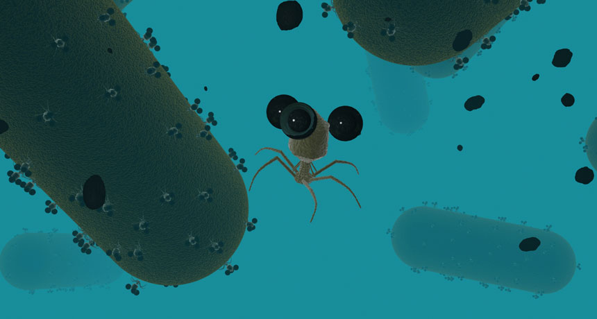
Phages identify and grab bacteria using proteins on their leglike tail fibers, which form a strong bond with compounds on the bacterial cell surface. To infect the cell, the phage injects its genetic material. This hijacks the cell, forcing its machinery to produce phage clones.
Nugen and collaborators programmed phages to tag E. coli bacteria. The team’s engineered phages contained extra DNA that told the bacteria to make an easily detectable enzyme. When the infection caused the bacterial cells to rupture and release the new phages, a chemical reaction involving the enzyme produced a measurable signal: light, color or an electric current. For example, the phages exposed E. coli in milk and orange juice by turning the liquids red or pink.
The researchers also loaded the phages with nanoparticles with a magnetic iron and cobalt core. Once the phages latched onto the bacteria, researchers could use a magnet to round the bacteria up even before the bacteria ruptured and announced their presence. This allowed the researchers to detect low concentrations of bacteria: less than 10 E. coli cells in half a cup of water. Conventional methods grow the bacteria into colonies to find them, which can take up to two days. But using the phages, Nugen and his colleagues skipped this step and found the cells within a few hours.
Using phages for magnetic separation would be “really nice for food and environmental samples because they tend to be really dirty,” said Michael Wiederoder, a bioengineer at the U.S. Army Natick Soldier Research, Development and Engineering Center in Massachusetts, who was not involved in the research. The salt, sugar and fats in food can slow the reactions of antibody-based tests, he said.
Also, the phages infect only bacteria that can reproduce, allowing testers to tell the difference between live cells and those killed by antibiotics, heat or chlorine. With food, “whether the bacteria are alive or dead is the difference between you getting sick and not,” Wiederoder said.
The nanobots could also prove useful for blood or other human samples. There, phages would provide a way to find resistant bacteria left alive after a course of antibiotics.
The next challenge: tinkering with the phages to tune which bacteria they go after. In nature, phages prey on specific species. But in food, it may be helpful to detect several common offenders, like E. coli, Salmonella and Listeria, or, alternatively, to have greater discrimination to find only the pathogenic E. coli and leave the rest.
When a stoned Carl Sagan used to do his TV bit and talk about billions and billions of galaxies, I turned my world inward, to the trillions and trillions of viruses.
I tell daughter Sorenne, I don’t care which you focus on, but get one of them right.
According to Jim Robbins of the New York Times,high in the Sierra Nevada mountains of Spain, an international team of researchers set out four buckets to gather a shower of viruses falling from the sky.
Scientists have surmised there is a stream of viruses circling the planet, above the planet’s weather systems but below the level of airline travel. Very little is known about this realm, and that’s why the number of deposited viruses stunned the team in Spain. Each day, they calculated, some 800 million viruses cascade onto every square meter of the planet.
Most of the globe-trotting viruses are swept into the air by sea spray, and lesser numbers arrive in dust storms.
“Unimpeded by friction with the surface of the Earth, you can travel great distances, and so intercontinental travel is quite easy” for viruses, said Curtis Suttle, a marine virologist at the University of British Columbia. “It wouldn’t be unusual to find things swept up in Africa being deposited in North America.”
The study by Dr. Suttle and his colleagues, published earlier this year in the International Society of Microbial Ecology Journal, was the first to count the number of viruses falling onto the planet. The research, though, is not designed to study influenza or other illnesses, but to get a better sense of the “virosphere,” the world of viruses on the planet.
Generally it’s assumed these viruses originate on the planet and are swept upward, but some researchers theorize that viruses actually may originate in the atmosphere. (There is a small group of researchers who believe viruses may even have come here from outer space, an idea known as panspermia.)
Whatever the case, viruses are the most abundant entities on the planet by far. While Dr. Suttle’s team found hundreds of millions of viruses in a square meter, they counted tens of millions of bacteria in the same space.
Mostly thought of as infectious agents, viruses are much more than that. It’s hard to overstate the central role that viruses play in the world: They’re essential to everything from our immune system to our gut microbiome, to the ecosystems on land and sea, to climate regulation and the evolution of all species. Viruses contain a vast diverse array of unknown genes — and spread them to other species.
Last year, three experts called for a new initiative to better understand viral ecology, especially as the planet changes. “Viruses modulate the function and evolution of all living things,” wrote Matthew B. Sullivan of Ohio State, Joshua Weitz of Georgia Tech, and Steven W. Wilhelm of the University of Tennessee. “But to what extent remains a mystery.”
We’re all hosts on a viral planet.
I didn’t understand this until fourth-year university, and it was only then I became interested in learning.
Until then, I was bored.
Researchers recently identified an ancient virus that inserted its DNA into the genomes of four-limbed animals that were human ancestors. That snippet of genetic code, called ARC, is part of the nervous system of modern humans and plays a role in human consciousness — nerve communication, memory formation and higher-order thinking. Between 40 percent and 80 percent of the human genome may be linked to ancient viral invasions.
Viruses and their prey are also big players in the world’s ecosystems. Much research now is aimed at factoring their processes into our understanding of how the planet works.
“If you could weigh all the living material in the oceans, 95 percent of it is stuff is you can’t see, and they are responsible for supplying half the oxygen on the planet,” Dr. Suttle said.
In laboratory experiments, he has filtered viruses out of seawater but left their prey, bacteria. When that happens, plankton in the water stop growing. That’s because when preying viruses infect and take out one species of microbe — they are very specific predators — they liberate nutrients in them, such as nitrogen, that feed other species of bacteria. In the same way, an elk killed by a wolf becomes food for ravens, coyotes and other species. As plankton grow, they take in carbon dioxide and create oxygen.
One study estimated that viruses in the ocean cause a trillion trillion infections every second, destroying some 20 percent of all bacterial cells in the sea daily.
Viruses help keep ecosystems in balance by changing the composition of microbial communities. As toxic algae blooms spread in the ocean, for example, they are brought to heel by a virus that attacks the algae and causes it to explode and die, ending the outbreak in as little as a day.
While some viruses and other organisms have evolved together and have achieved a kind of balance, an invasive virus can cause rapid, widespread changes and even lead to extinction.
It’s winter in Brisbane, Australia, with highs in the 90s F (30s C) a couple of weeks ago, and today where I went to the arena for a lunchtime skate with Amy in shorts and the loudest Hawaiian shirt I own (additional layers were added once in the arena), and where what they call gastro outbreaks have increased dramatically.
Emergency rooms throughout Brisbane have been overwhelmed, and not just by dumbass Canadians falling off bikes.
But what is a gastro bug?
How can they not name the bug?
Regis aged care facility in the suburb of Yeronga, just down the road from us, has been in lockdown for 26 days.
A Regis spokesperson on Tuesday night reiterated “there have been no deaths confirmed as being as a result of gastro.”
“As advised previously, Regis has experienced an episode of gastroenteritis at the Yeronga facility. It was first identified on 28 July. We are pleased to say that the episode is nearing completion.”
Darren Cartwright of the Courier-Mail reported yesterday there has been a four-fold increase in gastroenteritis outbreaks in Brisbane’s daycare centres, with almost 200 children alone affected on the southside since June.
In total more than 50 daycare centres have alerted Queensland Health of an outbreak of gastroenteritis.
A Queensland Health spokesman acknowledged the outbreaks were “significantly” higher this year than for the same eight week periods in 2016.
“The data indicates a significantly high number of outbreaks during this eight week period in 2017, however, it should be noted that half of these outbreaks involved fewer than 10 unwell children,” the spokesman said.
That will make the parents and kids feel better.
“In general, it has been a big year for viral gastroenteritis outbreaks across the region.”
At least, that’s what scientists thought a few days ago. Now a new study published Thursday is making researchers rethink how some viruses could infect animals.
A team at the U.S. Army Medical Research Institute of Infectious Diseases has found a mosquito virus that’s broken up into pieces. And the mosquito needs to catch several of the pieces to get an infection.
“It’s the most bizarre thing,” says Edward Holmes, a virologist at the University of Sydney, who wasn’t involved in the study. It’s like the virus is dismembered, he says.
“If you compare it to the human body, it’s like a person would have their legs, trunk and arms all in different places,” Holmes says. “Then all the pieces come together in some way to work as one single virus. I don’t think anything else in nature moves this way.”
Most viruses have simple architecture. They have a few genes — say about a half-dozen or so — that are packaged up into a little ball, 1/500th the width of a human hair.
“You can think of it like a teeny-weeny tennis ball with spikes,” Holmes says.
When the virus infects a cell, the ball latches onto the cell’s surface, opens up and pops its genes into the cell.
Poof! The cell is infected. That’s all it takes. One ball, sticking to one cell.
But that’s not the case for the Guaico Culex virus. It has five genes. And each one gets stuffed into a separate ball. Imagine five tennis balls, each with a different color: a red tennis ball, a blue one, a green one, a yellow one and an orange one.
Then to get infected with the virus, a mosquito needs to catch at least four different colored balls, researchers write in the journal Cell Host & Microbe. Otherwise the infection fails.
“The fifth ball seems to be optional,” says Jason Ladner, a genomicist at USAMRIID, who helped discover the virus. Getting the fifth one could control how dangerous the virus is, he says.
Ladner and his team found the virus inside a Culex mosquito found in Guaico, Trinidad — hence the name of the virus, Guaico Culex. Culex mosquitoes are common across the U.S. and spread West Nile Virus.
The study is part of a larger project aimed at figuring out what viruses, in addition to Zika and yellow fever, could be lurking inside mosquitoes and possibly waiting to spill over into people.
Indeed, each year, scientists are finding thousands of new viruses, says Vincent Racaniello, at Columbia University. “It’s hard to put a number on it. But it’s huge.”
“We finally have the tools to find them,” he says.
But that doesn’t mean we can immediately understand what they do, or even whom they infect.
“There’s so much we don’t know about viruses,” Racaniello adds. And with viruses, really anything is possible. “We should always expect the unexpected,” he says.
To assess the safety of Danube water for bathing, physical, chemical, bacteriological tests were performed. While many parameters for monitoring the quality of water are regulated by law, there are neither national nor international legislations addressing the presence of viruses in recreational waters. In this study, we performed analyses that surpassed national requirements, and investigated if adenovirus, enterovirus or rotavirus genetic material was present in samples of recreational water collected for quality monitoring.
Of 90 water samples obtained during the study, enterovirus material was not found in any sample, but adenovirus and rotavirus genetic materials were respectively detected in 60 and 31 samples. Statistical analyses showed a significant correlation between adenovirus DNA and total coliforms in the water. Even when water samples were adequate for recreational use, adenoviruses were detected in 75% (57/76) of such samples. Our results indicate that implementation of viral indicators in recreational water might be helpful to better assess public health safety. This might be particularly relevant in areas where urban wastewater treatment is insufficient and surface waters affected by wastewater are used for recreation.
Testing For Viral Material In Water Of Public Bathing Areas Of The Danube During Summer, Vojvodina, Serbia, 2014
Eurosurveillance, Volume 21, Issue 15, 14 April 2016
A Jovanović Galović, S Bijelović, V Milošević, I Hrnjaković Cvjetkovic, M Popović, G Kovačević , J Radovanov, N Dragić, V Petrović
In an effort to understand and eventually reduce the incidence of foodborne illnesses, University of Illinois researchers studied the ability of pathogenic viruses to adhere to fresh produce surfaces.
“We chose 24 of the most common salad vegetables in the U.S. and assayed them to see if there was any relationship between the morphology and chemistry of the leaf or fruit surface and the adherence of viral particles, before and after a washing treatment,” says U of I geneticist Jack Juvik.
The researchers inoculated leafy salad greens and tomatoes with a swine virus that mimics human rotavirus, a common pathogen responsible for diarrhea, vomiting, fever, and abdominal pain. After exposing the vegetable surfaces to the virus, the researchers rinsed the vegetables twice with a standard saline solution.
“We correlated virus adherence to roughness of the surface at different scales. We also looked at the chemistry of the proteins and waxes associated with the leaf cuticle – a waxy layer that protects the plant against diseases and reduces water loss,” Juvik explains. “Before this, no one had tested the relationship between chemistry and surface texture on the adherence of virus particles.”
The researchers found a thousand-fold difference in the number of viral particles adhering to different types of leafy greens and tomatoes. Vegetables with three-dimensional crystalline wax structures on the leaf cuticle harbored significantly fewer virus particles after rinsing. This was counterintuitive, as it was expected that small virus particles could “hide” in the rough structures of these cuticles.
“I was surprised, too,” Juvik says. “But normally, viruses adhere to oxygen groups, like OH, which are associated with proteins and carbohydrates on the surface. When the wax completely covers the surface, it becomes totally hydrophobic, which renders the whole leaf surface harder for viruses to attach to. Furthermore, rinsing those leaves with water gives the viruses the OH groups they’re looking for, so they’re easier to wash away.”
Produce is exposed to viruses and other pathogens in a number of ways, including contaminated irrigation water, animal wastes, and handling by sick workers. But because salad vegetables are consumed fresh, pathogens cannot be killed by cooking or most other sterilization methods.
“Viruses are literally everywhere, causing many opportunities for infection. But the information from this study can be used down the road to select or breed for varieties that might have the capacity to reduce adherence of these particles,” Juvik explains.
The researchers have already repeated the study using the bacterium E. coli, but they plan to look at even more vegetable varieties and pathogens in future studies.
The article, “Influence of epicuticular physiochemical properties on porcine rotavirus adsorption to 24 leafy green vegetables and tomatoes” was published in PLOS One. The study was led by Lu Lu, whose co-authors included Juvik, Kang-Mo Ku, Sindy Paola Palma-Salgado, Andrew Page Storm, Hao Feng, and Thanh Nguyen, all from the University of Illinois. The project received funding from the USDA’s National Institute of Food and Agriculture.