Salmonella Dublin is nasty

Salmonella enterica serotype Dublin is a cattle-adapted bacterium that typically causes bloodstream infections in humans.

To summarize demographic, clinical, and antimicrobial drug resistance characteristics of human infections with this organism in the United States, we analyzed data for 1968–2013 from 5 US surveillance systems.

During this period, the incidence rate for infection with Salmonella Dublin increased more than that for infection with other Salmonella. Data from 1 system (FoodNet) showed that a higher percentage of persons with Salmonella Dublin infection were hospitalized and died during 2005–2013 (78% hospitalized, 4.2% died) than during 1996-2004 (68% hospitalized, 2.7% died). Susceptibility data showed that a higher percentage of isolates were resistant to >7 classes of antimicrobial drugs during 2005–2013 (50.8%) than during 1996–2004 (2.4%).

Epidemiology of Salmonella enterica Serotype Dublin infections among humans, United State, 1968-2013

Emerging Infectious Diseases, vol. 23, no. 9, September 2017, R. Reid Harvey, Cindy R. Friedman, Stacy M. Crim, Michael Judd, Kelly A. Barrett, Beth Tolar, Jason P. Folster, Patricia M. Griffin, and Allison C. Brown

https://wwwnc.cdc.gov/eid/article/23/9/17-0136_article

Dehydration and salt toxicity? Cows dying in Saskatchewan

Ann Hui of The Globe and Mail reports that for decades, cattle farmers have sent their livestock to graze on the sprawling Shamrock pasture, about 80 kilometres south of Chaplin in southwest Saskatchewan. Shamrock is where Russ Coward, a fourth-generation cattle farmer, has for years raised nearly a quarter of his cattle. It’s the same place Mr. Coward’s father sent his cattle.

But some time between last Monday and Friday, the cows and calves at Shamrock began to die. It’s not known whether the deaths happened all at once or slowly over the course of the four days. But by the time the manager arrived on Friday afternoon, 200 of the approximately 680 cattle in a single field were dead.

The president of Shamrock Grazing Ltd., Glenn Straub, called Mr. Coward, who raced out to the field. He was met with a gruesome scene. “We seen a tragedy,” he said. “We simply seen a terrible sight.”

Other ranchers soon joined him, about 31 in total who have cows and calves there. “We all had the same feelings – how did this happen? How did this happen?”

Since Friday, provincial authorities as well as the local RCMP have been trying to piece together the mystery. The cause is still being determined, but the prevailing theory is dehydration and salt toxicity.

The area has been subject to drought in recent weeks, said Saskatchewan’s chief veterinary officer, Betty Althouse. Officials believe this may have led to evaporation at the water source, resulting in higher concentrations of salt in the water.

“An analogy would be someone shipwrecked in the ocean,” Dr. Althouse told reporters this week. “They’re thirsty, they’re craving water, so they’re going to drink the water. But ultimately the salt water will kill them.”

Many of the dead cows and calves were found clustered around one “dugout” in particular – the pools of collected rain and runoff where they drink. Investigators at the Western College of Veterinary Medicine in Saskatoon who were called in to assist have collected samples from the dugouts. Results are expected some time this week.

Reinfection of cattle with E. coli

Enterohaemorrhagic Escherichia coli (EHEC), like E. coli O157:H7 are frequently detected in bovine faecal samples at slaughter. Cattle do not show clinical symptoms upon infection, but for humans the consequences after consuming contaminated beef can be severe.

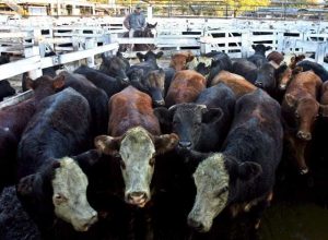
epa01322503 View of some of the more than 7,000 cattle that arrived to Liniers Market in Buenos Aires, Argentina, on 22 April 2008, to be slaughtered. Argentinean government and the biggest agriculture organizations had been discussing, this morning, the situation of milk sector that is one of the most conflictive topics on the negotiations with cow meat, grains and regional economies. EPA/CEZARO DE LUCA

The immune response against EHEC in cattle cannot always clear the infection as persistent colonization and shedding in infected animals over a period of months often occurs. In previous infection trials, we observed a primary immune response after infection which was unable to protect cattle from re-infection. These results may reflect a suppression of certain immune pathways, making cattle more prone to persistent colonization after re-infection.

To test this, RNA-Seq was used for transcriptome analysis of recto-anal junction tissue and ileal Peyer’s patches in nine Holstein-Friesian calves in response to a primary and secondary Escherichia coli O157:H7 infection with the Shiga toxin (Stx) negative NCTC12900 strain. Non-infected calves served as controls.

In tissue of the recto-anal junction, only 15 genes were found to be significantly affected by a first infection compared to 1159 genes in the ileal Peyer’s patches. Whereas, re-infection significantly changed the expression of 10 and 17 genes in the recto-anal junction tissue and the Peyer’s patches, respectively. A significant downregulation of 69 immunostimulatory genes and a significant upregulation of seven immune suppressing genes was observed.

Although the recto-anal junction is a major site of colonization, this area does not seem to be modulated upon infection to the same extent as ileal Peyer’s patches as the changes in gene expression were remarkably higher in the ileal Peyer’s patches than in the recto-anal junction during a primary but not a secondary infection. We can conclude that the main effect on the transcriptome was immunosuppression by E. coli O157:H7 (Stx) due to an upregulation of immune suppressive effects (7/12 genes) or a downregulation of immunostimulatory effects (69/94 genes) in the ileal Peyer’s patches. These data might indicate that a primary infection promotes a re-infection with EHEC by suppressing the immune function.

Potential immunosuppressive effects of Escherichia coli O157:H7 experimental infection on the bovine host

BMC Genomics; 2016; 17:1049; DOI: 10.1186/s12864-016-3374-y; Published: 21 December 2016

E. Kieckens, J. Rybarczyk, R. W. Li, D. Vanrompay, and E. Cox

http://bmcgenomics.biomedcentral.com/articles/10.1186/s12864-016-3374-y

Lower the loads: Phages don’t seem to work to lower E. coli O157 in cattle

Image

Escherichia coli O157:H7 is a major food safety concern for the beef industry. Several studies have provided evidence that cattle hides are the main source of beef carcass contamination during processing and that reductions in the E. coli O157:H7 load on the hides of cattle entering processing facilities will lead to reductions in carcass contamination. Bacteriophages have been proposed as a novel preharvest antimicrobial intervention to reduce the levels of E. coli O157:H7 on cattle hides.

lairageThe objective of this study was to evaluate a commercialized phage application administered in the lairage area of commercial beef processing plants for the ability to reduce E. coli O157:H7 contamination of cattle hides and carcasses. Cattle lots either received phage spray treatment (n = 289) or did not (n = 301), as they entered the lairage environments in two separate experiments at two different commercial beef processing plants. Hide and carcass samples were collected and analyzed for E. coli O157:H7 prevalence and concentration. Cattle hides receiving phage treatment had an E. coli O157:H7 prevalence of 51.8%, whereas untreated hides had a prevalence of 57.6%. For carcass samples, the E. coli O157 prevalence in treated and untreated samples was 17.1% and 17.6%, respectively.

The results obtained from these experiments demonstrated that the treatment of cattle hides with bacteriophages before processing did not produce a significant reduction of E. coli O157:H7 on cattle hides or beef carcasses during processing.

Evaluation of bacteriophage application to cattle in lairage at beef processing plants to reduce Escherichia coli O157:H7 prevalence on hides and carcasses

Arthur Terrance M., Kalchayanand Norasak, Agga Getahun E., Wheeler Tommy L., and Koohmaraie Mohammad

http://online.liebertpub.com/doi/abs/10.1089/fpd.2016.2189

Toddler contracts serious E. coli infection on NZ family farm

Eight months on from a rescue helicopter dash to Starship children’s hospital, two-year-old Grace Dheda is enjoying being back on her family’s farm – even though it nearly killed her.

grace-dhedaIn March, Grace and her family were savouring rural life in Wellsford.

Mum Megan and Dad Kirin were planning their up-coming wedding. 

That all came to a sudden halt when their daughter began to show signs of illness.

After two days of vomiting and diarrhea, a doctor diagnosed a tummy bug.

Grace was sent home and prescribed plenty of fluids, Megan says.

At home Grace played on the deck like her normal self, but collapsed at bedtime.

Grace was rushed back to the doctors.

“They put her on oxygen straight away. She’d been unconscious for about 45 minutes and they were starting to worry about potential brain damage.”

Given the severity of the situation and the closest ambulance an hour away, the Auckland Westpac Rescue Helicopter was called.

Grace and Megan were ferried to a helipad and arrived to see the chopper landing.

“It was such a relief to see the helicopter,” Megan says.

Megan recalls, “At first nobody knew what was wrong with her and why she was having these seizures. It wasn’t until a few days before we left the hospital that we found out she had contracted E. coli and HUS (Hemolytic Uremic Syndrome).”

HUS is a severe complication of the E. coli infection that can lead to kidney failure.

At first it was thought that Grace had contracted the bacterial infection through the water supply, however this was later tested and found to be normal.

It is now believed that she contracted it via the farm animals.

Megan says, “We’ve got cows here on the farm and I don’t like Grace going anywhere near them. The doctor told me I have ‘parental anxiety.’ ‘I love the farm life, but I’m a bit paranoid now and have about 20 bottles of sanitiser around the place.”

The Helicopter Trust is actively fundraising at present in order to purchase three new ventilators for use on their helicopters and in their Rapid Response Vehicle.

A lot of STEC: Numbers mean new interventions

Shiga toxin-producing Escherichia coli (STEC) strains can colonize cattle for several months and may, thus, serve as gene reservoirs for the genesis of highly virulent zoonotic enterohemorrhagic E. coli (EHEC).

Cattle-Heat-Load-ForecastingAttempts to reduce the human risk for acquiring EHEC infections should include strategies to control such STEC strains persisting in cattle. We therefore aimed to identify genetic patterns associated with the STEC colonization type in the bovine host. We included 88 persistent colonizing STEC (STECper) (shedding for ≥4 months) and 74 sporadically colonizing STEC (STECspo) (shedding for ≤2 months) isolates from cattle and 16 bovine STEC isolates with unknown colonization types. Genoserotypes and multilocus sequence types (MLSTs) were determined, and the isolates were probed with a DNA microarray for virulence-associated genes (VAGs). All STECper isolates belonged to only four genoserotypes (O26:H11, O156:H25, O165:H25, O182:H25), which formed three genetic clusters (ST21/396/1705, ST300/688, ST119). In contrast, STECspo isolates were scattered among 28 genoserotypes and 30 MLSTs, with O157:H7 (ST11) and O6:H49 (ST1079) being the most prevalent. The microarray analysis identified 139 unique gene patterns that clustered with the genoserotypes and MLSTs of the strains. While the STECper isolates possessed heterogeneous phylogenetic backgrounds, the accessory genome clustered these isolates together, separating them from the STECspo isolates.

Given the vast genetic heterogeneity of bovine STEC strains, defining the genetic patterns distinguishing STECper from STECspo isolates will facilitate the targeted design of new intervention strategies to counteract these zoonotic pathogens at the farm level.

The accessory genome of Shiga toxin-producing Escherichia coli defines a persistent colonization type in cattle

Appl Environ Microbiol 82:5455–5464. doi:10.1128/AEM.00909-16.

SA Barth, C Menge, I Eichhorn, T Semmier, LH Wieler, D Pickard, A Belka, C Berens, L Geue

http://aem.asm.org/content/82/17/5455.abstract?etoc

Not a Letterman list: Top 6 STECs in Canada

The incidence of the top 6 STEC serotypes was determined in two beef slaughter houses. In total, 328 samples were taken of hides, de-hided carcasses and the plant environment. Samples were enriched in Tryptic Soy Broth containing novobiocin then screened using RT-PCR GeneDiskÒ system that targeted stx, eae and wzx genes.

top.10.lettermanIt was found that 92.5% (172 of 186) of the hide samples. 72.5% (29 of 40) de-hided samples and 84.3% (86 of 102) of the environmental samples returned presumptive positive results. Serotypes O103, O45 and O121 were most commonly encountered although all the Top 6 serotypes were represented within individual samples. However, attempts to recover the Top 6 serotypes by culturing proved unsuccessful despite screening up to 20 colonies per CHROMAgar® plate of enriched sample. The reasons for the discrepancy between the RT-PCR and culture methods were found to be due to low levels of the target in enriched samples, presence of virulence factors in different cells and also the transient retention of stx. With regards the latter it was found that strains harboring a full set of virulence factors (eae, stx) were more common in grown cultures held post-incubation at 4 °C for 14 days. Moreover, no stx gene was recovered when isolates were sub-cultured on TSA but was present in the same strains grown on CHROMAgar®. In total 39 STEC isolates were recovered with the majority harboring stx1, stx2, eae and hylA. Only 3 of the isolates had stable complement of virulence factors and were identified as O172:H28, O76:H7 and O187:H52.

Although no Top 6 STEC were isolated the presence of virulent strains on carcasses with the potential to cause Hemolytic Uremic Syndrome is of concern. The significance of those STEC that transiently harbor virulence factors is unclear although clearly impacts on diagnostic performance robustness when screening for the Top 6 non-O157 STEC.

Incidence of Top 6 shiga toxigenic Escherichia coli within two Ontario beef processing facilities: Challenges in screening and confirmation testing

AIMS Microbiology, 2016, 2(3): 278-291. DOI: 10.3934/microbial.2016.3.278

Bannon, M. Melebari, C. Jordao Jr., C.G. Leon-Velarde, K. Warriner

http://www.aimspress.com/article/10.3934/microbiol.2016.3.278

Cattle be shedding STECs

Shiga toxin-producing Escherichia coli (STEC) is an important foodborne pathogen that can cause hemorrhagic colitis and hemolytic-uremic syndrome. Cattle are the primary reservoir for STEC, and food or water contaminated with cattle feces is the most common source of infections in humans.

beef.cattleConsequently, we conducted a cross-sectional study of 1,096 cattle in six dairy herds (n = 718 animals) and five beef herds (n = 378 animals) in the summers of 2011 and 2012 to identify epidemiological factors associated with shedding.

Fecal samples were obtained from each animal and cultured for STEC. Multivariate analyses were performed to identify risk factors associated with STEC positivity. The prevalence of STEC was higher in beef cattle (21%) than dairy cattle (13%) (odds ratio [OR], 1.76; 95% confidence interval [CI], 1.25, 2.47), with considerable variation occurring across herds (range, 6% to 54%). Dairy cattle were significantly more likely to shed STEC when the average temperature was >28.9°C 1 to 5 days prior to sampling (OR, 2.5; 95% CI, 1.25, 4.91), during their first lactation (OR, 1.8; 95% CI, 1.1, 2.8), and when they were <30 days in milk (OR, 3.9; 95% CI, 2.1, 7.2). These data suggest that the stress or the negative energy balance associated with lactation may result in increased STEC shedding frequencies in Michigan during the warm summer months.

Future prevention strategies aimed at reducing stress during lactation or isolating high-risk animals could be implemented to reduce herd-level shedding levels and avoid transmission of STEC to susceptible animals and people.

STEC shedding frequencies vary considerably across cattle herds in Michigan, and the shedding frequency of strains belonging to non-O157 serotypes far exceeds the shedding frequency of O157 strains, which is congruent with human infections in the state. Dairy cattle sampled at higher temperatures, in their first lactation, and early in the milk production stage were significantly more likely to shed STEC, which could be due to stress or a negative energy balance. Future studies should focus on the isolation of high-risk animals to decrease herd shedding levels and the potential for contamination of the food supply.

Factors associated with Shiga toxin-producing Escherichia coli shedding by dairy and beef cattle

Cristina Venegas-Vargasa*, Scott Hendersona,b, Akanksha Khareb*,Rebekah E. Moscib, Jonathan D. Lehnertb*, Pallavi Singhb,Lindsey M. Ouelletteb*, Bo Norbya, Julie A. Funka, Steven Rustc, Paul C. Bartletta,Daniel Groomsa and Shannon D. Manningb

aDepartment of Large Animal Clinical Sciences, College of Veterinary Medicine, Michigan State University, East Lansing, Michigan, USA

bDepartment of Microbiology and Molecular Genetics, Michigan State University, East Lansing, Michigan, USA

cDepartment of Animal Science, Michigan State University, East Lansing, Michigan, USA

Applied and Environmental Microbiology, August 2016, Volume 82, Number 16, Pages 5049-5056, doi:10.1128/AEM.00829-16

http://aem.asm.org/content/82/16/5049.abstract?etoc

E. coli O165:H25 in Nebraska cattle

Russ Rice a DVM from Broken Bow, Nebraska, writes in Bovine Veterinarian that a recent experience in a Nebraska feedlot, supported by extensive diagnostic work from veterinarians at the University of Nebraska-Lincoln, indicates Escherichia coli O165:H25, an enterohemorrhagic strain, can cause disease in cattle and potentially poses a food-safety hazard for humans.

Russ RiceNebraska veterinarians whose work on this case proved critical in establishing a diagnosis and analyzing the implications included Bruce Brodersen, DVM, PhD, Dee Griffin, DVM, MS, John D. Loy, DVM, PhD and Rodney A. Moxley, DVM, PhD.

The case report was published in JMM Case Reports in November 2015, in an article titled “Haemorrhagic colitis associated with enterohaemorrhagic Escherichia coli O165:H25 infection in a yearling feedlot heifer.” Authors, in addition to Brodersen, Loy and Moxley, included Zachary R. Stromberg and Gentry L. Lewis from the University of Nebraska School of Veterinary Medicine and Biomedical Sciences, and Isha R. Patel and Jayanthi Gangiredla from the FDA’s Center for Food Safety and Applied Nutrition.

This case began in early January, 2014, with a pen of 170 Iowa-origin heifers, averaging 160 pounds, in a Nebraska feedlot. The 54 mega-calorie ration included dry and high moisture corn, 32% wet distillers’ grains, syrup, roughage, standard supplements and Bovatec.

In February, the feedlot crew began pulling some heifers from the pen for respiratory disease treatment. Crews noted that some bloody stools were present in the pen, and we treated the cattle with amprolium based on a presumptive diagnosis of coccidiosis. Bloody stools persisted though, and one animal died and another showed neurological signs and bloody diarrhea on February 18th. We euthanized that heifer using methods approved by the University of Nebraska Institutional Animal Care and Use Committee, and conducted a necropsy immediately. The gross lesion appeared to be acute inflammation of the cecum and colon. Our differential diagnosis included coccidiosis, salmonellosis, BVD, bovine coronavirus and clostridial enteritis.

We prepared fresh and fixed specimens and sent them to the UNL diagnostic laboratory for analysis. Dr. Loy’s laboratory isolated a strain of E. coli from the colonic mucosal tissue, and Dr. Brodersen noted histopathologic lesions in the colon that were suggestive of infection with a strain of attaching-and-effacing E. coli. He consulted with Dr. Moxley, an E. coli researcher, who requested the gross and fixed tissues for further study. Upon close examination, Dr. Moxley found small erosions in the colonic mucosa which he photographed.  He then had his laboratory conduct other cultures, and had the original E. coli isolate serotyped.  The isolate was determined to be an O165:H25 serotype.  At this point, Dr. Moxley obtained specific antiserum to O165, and had the histology laboratory of the diagnostic lab conduct immunohistochemistry using this antiserum. Dr. Moxley found O165-positive bacteria in lesion sites corresponding to the gross erosions, and it was then that the cause of the lesions was confirmed. He had the isolate tested further by microarray analysis through collaborators at the FDA. It was found to contain virulence genes corresponding to type III secretion system (T3SS) structure and regulation. Microarray analysis of another E. coli strain (serotype O145:H28) isolated from the same tissue by the Moxley laboratory ruled the latter organism out as a pathogen in the case because that strain was found to have a number of mutations that inactivated virulence genes, such as those of the T3SS. Details of the molecular typing are included in the paper published in JMM Case Reports. …

The O165:H25 serotype is similar to E. coli O157:H7, and could be an emerging food-borne pathogen in cattle and beef. The pathogen has been isolated from the feces and beef carcasses of cattle, but has not previously been associated with disease in cattle. UNL scientists note that the serotype has been involved in uncommon, but potentially severe disease in humans. Based on findings in this case, we suggest veterinarians include enterohemorrhagic E. coli in their list of differential diagnoses when they see enteric disease, particularly bloody diarrhea, in feedlot cattle. If EHEC is suspected, veterinarians should work with their diagnostic laboratory to ensure proper collection, handling and submission of appropriate samples for diagnosis.

At the diagnostic laboratory, Drs. Moxley, Brodersen, and Loy indicate that these lesions might not have been seen in samples taken as early as ten minutes of death. Fortunately in this case, in which we euthanized the heifer and were able to collect samples immediately, we were able to verify this unexpected diagnosis. Most of the time, post-mortem changes will occur before we get the opportunity to collect samples.

Several veterinarians have commented to me they may have missed cases of EHEC in the past. I probably have missed these as well. Going forward, I plan to take extra care in collecting samples and communicating this to our clients. This case serves as a good reminder of the public-health risk we face on a daily basis. We all need to emphasize this to our clients and staff.
From Promed: E. coli O157 in Africa, 1992

The Epicore Global Surveillance Project reports that in 1992, a large outbreak of bloody diarrhea caused by Escherichia coli O157 infections occurred in southern Africa.

Swaziland-Bucket-BrigadeIn Swaziland, 40,912 physician visits for diarrhea in persons aged 5 years and older were reported during October through November 1992. This was a 7-fold increase over the same period during 1990-91. The attack rate was 42 percent among 778 residents we surveyed. Female gender and consuming beef and untreated water were significant risks for illness.

E. coli O157:NM was recovered from 7 affected foci in Swaziland and South Africa; 27 of 31 patients and environmental isolates had indistinguishable pulsed-field gel electrophoresis patterns. Compared with previous years, a 5-fold increase in cattle deaths occurred in October 1992. The 1st heavy rains fell that same month (36 mm), following 3 months of drought. Droughts, carriage of E. coli O157 by cattle, and heavy rains with contamination of surface water appear to be important factors contributing to this outbreak.

Early in November 1992, physicians arriving for duty at a small hospital on a sugar plantation in Swaziland found over 100 persons sprawled on the ground in front of the casualty department. Many had bloody diarrhea, and almost all were suffering severe abdominal pains. The next day, the number of patients with the dysenteric illness nearly doubled, yet stool specimens sent to local laboratories did not yield common parasitic or bacterial pathogens, including Shigella spp. With the etiologic agent still unknown into the 2nd week of the outbreak, specimens were forwarded to a reference laboratory in South Africa, where a surprising discovery was made: E. coli O157 had emerged in Africa.

An outbreak of E. coli O157 infections was heretofore unheard of in Africa or, for that matter, anywhere in the developing world. E. coli O157 had been isolated only once before in southern Africa, from an elderly man undergoing surgery for lower gastrointestinal bleeding in Johannesburg in 1990. Carriage of E. coli O157 by cattle, cattle deaths secondary to drought, and heavy rains that resulted in contamination of surface water were important factors contributing to the emergence of E. coli O157 in Africa. Given that drought and heavy rains will likely recur in Africa, the possibility that E. coli O157 will once again emerge to cause a major regional outbreak cannot be excluded. Clinicians need to be aware of this so that delayed diagnosis and inappropriate treatment resulting in loss of lives can be avoided.