Going down, in the sink

Since knowledge and understanding of waterborne pathogens and their diseases are well illuminated, a few research publications on the prevalence of pathogenic microorganisms in various household sink drain pipes are often not extensively examined. Therefore, this study aims to (a) assess and monitor the densities of the bacterial community in the different natural biofilm that grow on plastic pipelines, (b) to detect Escherichia coli , Salmonella , and Listeria spp. from natural biofilm samples that are collected from the kitchen (n = 30), bathroom (n = 10), laboratories (n = 13), and hospital (n = 8) sink drainage pipes.

Three bacterial species selected were assessed using a culture‐dependent approach followed by verification of isolates using both BIOLOG GEN III and polymerase chain reaction. The estimated number of each bacterium was 122 isolates, while 60, 20, 26, and 16 isolates were obtained from the natural biofilm samples, kitchen, bathroom, laboratories, and hospital, respectively. As for the tests, in all types of biofilm samples, the overall bacterial counts at low temperature (22°C) were higher than those at high temperature (37°C). Meanwhile, E . coli had the most significant number of bacterial microorganisms compared to the other two pathogens. Additionally, the most massive cell densities of E . coli , Salmonella , and Listeria species were discovered in the biofilm collected from the kitchen, then the hospital.

Statistically, the results reveal that there is a positive correlation (p ≥ .0001) with significance between the sources of biofilm. This work certainly makes the potential of household sink drain pipes for reservoir contagious pathogens more explicitly noticeable. Such knowledge would also be beneficial for prospective consideration of the threat to human public health and the environment.

Prevalence of E. coli, salmonella, and listeria spp. as potential pathogens: A comparative study for biofilm of sink drain environment

Journal of Food Safety

Mohamed Azab El‐Liethy, Bahaa A. Hemdan, Gamila E. El‐Taweel

https://doi.org/10.1111/jfs.12816

https://onlinelibrary.wiley.com/doi/abs/10.1111/jfs.12816?af=R

Foodborne pathogen sheltered by harmless bacteria that support biofilm formation

Pathogenic bacteria that stubbornly lurk in some apple-packing facilities may be sheltered and protected by harmless bacteria that are known for their ability to form biofilms, according to Penn State researchers, who suggest the discovery could lead to development of alternative foodborne-pathogen-control strategies. 

That was the key finding that emerged from a study of three tree-fruit-packing facilities in the Northeast where contamination with Listeria monocytogenes was a concern. The research, done in collaboration with the apple industry, was an effort to better understand the microbial ecology of food-processing facilities. The ultimate goal is to identify ways to improve pathogen control in the apple supply chain to avoid foodborne disease outbreaks and recalls of apples and apple products.

“This work is part of Penn State’s efforts to help producers comply with standards set forth in the federal Food Safety Modernization Act, often referred to as FSMA,” said researcher Jasna Kovac, assistant professor of food science, College of Agricultural Sciences. “The Department of Food Science at Penn State, through research and extension activities, has an ongoing collaboration with the apple industry, led by Luke LaBorde, professor of food science.”

The research was done in collaboration with the apple industry, in an effort to better understand the microbial ecology of food-processing facilities. The ultimate goal is to identify ways to improve pathogen control in the apple supply chain to avoid foodborne disease outbreaks and recalls of apples and apple products. 

In the study, researchers sought to understand the composition of microbiota in apple-packing environments and its association with the occurrence of the foodborne pathogen Listeria monocytogenes. Their testing revealed that a packing plant with a significantly higher Listeria monocytogenes occurrence was uniquely dominated by the bacterial family Pseudomonadaceae and the fungal family Dipodascaceae.

“As we investigated the properties of these microorganisms, we learned that they are known to be very good biofilm formers,” said lead researcher Xiaoqing Tan (upper left), a recently graduated master’s degree student in food science and a member of the Penn State Microbiome Center, housed in the Huck Institutes of the Life Sciences. “Based on our findings, we hypothesize that these harmless microorganisms are supporting the persistence of Listeria monocytogenes because they protect the harmful bacteria by enclosing them in biofilms. We are testing this hypothesis in a follow-up study.”

The findings of the research, published today (Aug. 21) in Microbiome, provide insight into the Listeria contamination problem and may lead to researchers and the apple industry getting closer to solving it, Kovac believes. Equipment in fruit-processing plants — such as brush conveyors — have a poor sanitary design that makes them difficult to clean and sanitize, she pointed out. She and LaBorde plan to work with the apple industry to devise more effective cleaning and sanitizing strategies.

Camera 360

Researchers collected samples in apple-packing facilities in which Listeria monocytogenes has been persistent. They discovered that harmless bacteria may be sheltering the pathogens.

 “Following up on these findings, we are experimenting with some of the nonpathogenic strains of bacteria that are not harmful to humans to see whether they can be used as biocontrols,” she said. “Once applied on the surfaces of the equipment in these environments, they may be able to outcompete and suppress Listeria, thus reducing food-safety risks and potential regulatory action. We are still exploring that approach in a controlled laboratory environment. If it proves to be feasible, we would like to test it in apple-packing and processing facilities.”

The challenge presented by microbiota possibly sheltering Listeria monocytogenes is not limited to fruit-processing facilities or produce, Penn State researchers suspect. They will soon begin analyzing microbial communities in dairy-processing facilities to determine the microbial composition and ecology of these environments.

Biofilms be good protection for bugs

In nature and man-made environments, microorganisms reside in mixed-species biofilms, in which the growth and metabolism of an organism are different from these behaviors in single-species biofilms. Pathogenic microorganisms may be protected against adverse treatments in mixed-species biofilms, leading to health risk for humans. Here, we developed two mixed five-species biofilms that included one or the other of the foodborne pathogens Listeria monocytogenes and Staphylococcus aureus.

The five species, including the pathogen, were isolated from a single food-processing environmental sample, thus mimicking the environmental community. In mature mixed five-species biofilms on stainless steel, the two pathogens remained at a constant level of ∼105 CFU/cm2. The mixed five-species biofilms as well as the pathogens in monospecies biofilms were exposed to biocides to determine any pathogen-protective effect of the mixed biofilm. Both pathogens and their associate microbial communities were reduced by peracetic acid treatments. S. aureus decreased by 4.6 log cycles in monospecies biofilms, but the pathogen was protected in the five-species biofilm and decreased by only 1.1 log cycles. Sessile cells of L. monocytogenes were affected to the same extent when in a monobiofilm or as a member of the mixed-species biofilm, decreasing by 3 log cycles when exposed to 0.0375% peracetic acid. When the pathogen was exchanged in each associated microbial community, S. aureus was eradicated, while there was no significant effect of the biocide on L. monocytogenes or the mixed community. This indicates that particular members or associations in the community offered the protective effect. Further studies are needed to clarify the mechanisms of biocide protection and to identify the species playing the protective role in microbial communities of biofilms.

IMPORTANCE This study demonstrates that foodborne pathogens can be established in mixed-species biofilms and that this can protect them from biocide action. The protection is not due to specific characteristics of the pathogen, here S. aureus and L. monocytogenes, but likely caused by specific members or associations in the mixed-species biofilm. Biocide treatment and resistance are a challenge for many industries, and biocide efficacy should be tested on microorganisms growing in biofilms, preferably mixed systems, mimicking the application environment.

Behavior of foodborne pathogens listeria monocytogenes and staphylococcus aureus in mixed-species biofilms exposed to biocides

Applied and Environmental Microbiology; DOI: 10.1128/AEM.02038-18

Virginie Oxaran, Karen Kiesbye Dittmann, et al

https://aem.asm.org/content/84/24/e02038-18?etoc=

Still need a better descriptor: Cross-contamination and cutting boards

Cross-contamination is one of the main factors related to foodborne outbreaks. This study aimed to analyze the cross-contamination process of Salmonella enterica serovar Enteritidis from poultry to cucumbers, on various cutting board surfaces (plastic, wood, and glass) before and after washing and in the presence and absence of biofilm.

Thus, 10 strains of Salmonella Enteritidis were used to test cross-contamination from poultry to the cutting boards and from thereon to cucumbers. Moreover, these strains were evaluated as to their capacity to form biofilm on hydrophobic (wood and plastic) and hydrophilic materials (glass).

We recovered the 10 isolates from all unwashed boards and from all cucumbers that had contacted them. After washing, the recovery ranged from 10% to 100%, depending on the board material. In the presence of biofilm, the recovery of salmonellae was 100%, even after washing. Biofilm formation occurred more on wood (60%) and plastic (40%) than glass (10%) boards, demonstrating that bacteria adhered more to a hydrophobic material.

It was concluded that the cutting boards represent a critical point in cross-contamination, particularly in the presence of biofilm. Salmonella Enteritidis was able to form a biofilm on these three types of cutting boards but glass showed the least formation.

Cross-Contamination and Biofilm Formation by Salmonella enterica Serovar Enteritidis on Various Cutting Boards

01.feb.18

Foodborne Pathogens and Deases, Volume 15, No. 2

Dantas Stéfani T. A. , Rossi Bruna F. , Bonsaglia Erika C. R. , Castilho Ivana G. , Hernandes Rodrigo T. , Fernandes Ary Júnior, and Rall Vera L. M.

https://doi.org/10.1089/fpd.2017.2341

It was probably the kitchen sink: 82 sick with Salmonella from UK restaurant 2015-16

From Eurosurveillance:

It is estimated that over 38,000 community cases of salmonellosis occur annually within the United Kingdom (UK) [1,2]. Salmonellosis often results from consumption of contaminated food or water [3], however, transmission via asymptomatic shedding by food handlers and exposure to contaminated environments where conditions are favourable for pathogen survival have also been implicated [3,4]. Here we report the findings of an investigation of an outbreak of salmonellosis where the environment was pivotal in continued transmission.

On 7 March 2015, Public Health England (PHE) East Midlands was alerted by the clinical microbiology laboratory of a local hospital to 21 cases of Salmonella enterica serovar Typhimurium gastroenteritis, with onset in February 2015. Seven cases in this initial phase of the outbreak required hospitalisation. Following this notification we suspected there was a community outbreak of S. Typhimurium; investigations and attempts to control the outbreak followed.

Hypothesis-generating interviews at the outset of the investigation identified that several cases had eaten at the same restaurant during the incubation period for their illness. Descriptive epidemiological analyses including subsequent cases pointed to the restaurant being the likely source. This popular, purpose (newly) built restaurant had opened only 18 months before the outbreak. The restaurant offered a full table-service menu, self-service salad bar and hot self-service carvery buffet serving roasted meats (turkey, beef, gammon and pork at weekends) and vegetables and condiments. Despite interventions to control the initial outbreak, cases continued to emerge followed by a prolonged period of transmission until 2016. The evolution of the investigation into this community outbreak and subsequent control measures is described, with specific reference to the use of whole genome sequencing (WGS) to link isolates and the role of the drains in continued pathogen transmission.

Mapping and visual inspection of the drainage systems identified significant issues. Water filled traps (u-bends) designed to prevent foul air flow from the drainage system into the building had failed and smoke testing revealed some ineffective drain seals, potentially allowing contaminated bio-aerosol to be disseminated into the kitchen. One sink drain was not connected to any drainage system with waste water pooling under the floor. Other larger drains had failed after leaking waste-water washed away the supporting substrate forming a cavity under the kitchen area. It transpired at that point that drainage water had, on occasion, risen into the kitchen area, although this had not been previously reported. Substantial remedial works were undertaken, however, these were found to have failed on re-inspection and so these drains were later decommissioned.

Biofilm [15] and flooded areas in underfloor cavities may have sustained this outbreak, after repeated environmental cleaning failed. Drainage problems in one area of the kitchen led to liquid from the drains seeping into the kitchen suggesting a contamination pathway. We found isolates matching the outbreak strain on kitchen cloths, swabs from kitchen sinks, and pot wash areas suggesting contact with sinks may have provided a second contamination pathway. We also identified ineffective drain water-traps potentially allowing the movement of contaminated bio-aerosols [13]. Smoke tests demonstrated the potential for dissemination of foul air into the kitchen.

Investigation using whole genome sequencing of a prolonged restaurant outbreak of salmonella typhimurium linked to the building drainage system, England, February 2015 to March 2016

Eurosurveillance, John Mair-JenkinsRoberta Borges-StewartCaroline HarbourJudith Cox-RogersTim Dallman, Philip AshtonRobert JohnstonDeborah ModhaPhilip MonkRichard Puleston,  https://doi.org/10.2807/1560-7917.ES.2017.22.49.17-00037

http://www.eurosurveillance.org/content/10.2807/1560-7917.ES.2017.22.49.17-00037


Polysorbate as possible E. coli food poisoning fix

Chris Waters, an associate professor of microbiology and molecular genetics at Michigan State University and colleauges report in the journal Biofouling that polysorbate, a safe additive found in everything from ice cream to cosmetics, seems to slow the toxic effects of E. coli poisoning.

alfalfa-sprouts-featuredThe findings show that polysorbates attack the protective biofilm in which E. coli lives and renders the deadly bacteria harmless.

“Biofilms are multicellular communities of bacteria that are usually encased in a protective slime,” says Waters. “We found that polysorbate 80 obliterates the biofilm and takes away the E. coli’s ability to damage the host during infection. We think this is due to blocking the ability of E. coli to produce toxin.”

Specifically, the team focused on the potent strain isolated from Germany that swept through Europe in 2011, causing thousands of infections and more than 50 deaths. Waters and Shannon Manning have previously studies this strain. Having samples of the bacteria at hand helped the team, led by Rudolph Sloup, a graduate student in microbiology and molecular genetics, isolate compounds that inhibited biofilms.

However, the results didn’t come easily. Waters and his team scoured scientific literature to identify anti-biofilm compounds, but none of them inhibited biofilms of this E. coli strain. Finally, the team found that the 20th compound tested, polysorbate 80, obliterated E. coli’s ability to form biofilms in the lab.

The next step was to determine if the compound was effective in an animal model of the disease by administering polysorbate 80 to infected mice in their drinking water.

“During our animal infection studies, polysorbate 80 had no effect on the numbers of infecting E. coli. This was a little shocking, especially based on how promising our earlier tests had been,” Waters says. “Later, though, our pathology tests showed that polysorbate 80 essentially blocked all toxicity, even though it didn’t reduce the number of bacteria.”

“Antibiotic use can often cause more harm than good with these types of E. coli infections because it causes the bacteria to release more toxin and it drives antimicrobial resistance,” Waters says. “Our results indicate that polysorbate 80 makes this strain of E. coli harmless, without these negative side effects. This approach also doesn’t disrupt patients’ natural microbiome leading to a healthier gut.”

Since polysorbate 80 is categorized as a GRAS (generally regarded as safe) compound, it doesn’t require FDA approval to be used as a treatment. Along with its potential for disarming the deadly German E. coli outbreak, polysorbate 80 could potentially help tackle more-common E. coli infections such as traveler’s diarrhea.

The next steps for this research will be to identify how polysorbate 80 inhibits biofilm formation and test its activity in other infection models.

Additional researchers from Michigan State and the University of Texas contributed to the study. Partial funding came from the National Institutes of Health and a Strategic Partnership Grant from the MSU Foundation.

Polysorbates prevent biofilm formation and pathogenesis of Escherichia coli O104:H4

The Journal of Bioadhesion and Biofilm Reseach, Volume 32, Issue 9, http://dx.doi.org/10.1080/08927014.2016.1230849

http://www.tandfonline.com/doi/abs/10.1080/08927014.2016.1230849?journalCode=gbif20&

Escherichia coli biotype O104:H4 recently caused the deadliest E. coli outbreak ever reported. Based on prior results, it was hypothesized that compounds inhibiting biofilm formation by O104:H4 would reduce its pathogenesis. The nonionic surfactants polysorbate 80 (PS80) and polysorbate 20 (PS20) were found to reduce biofilms by ≥ 90% at submicromolar concentrations and elicited nearly complete dispersal of preformed biofilms. PS80 did not significantly impact in vivo colonization in a mouse infection model; however, mice treated with PS80 exhibited almost no intestinal inflammation or tissue damage while untreated mice exhibited robust pathology. As PS20 and PS80 are classified as ‘Generally Recognized as Safe’ (GRAS) compounds by the Food and Drug Administration (FDA), these compounds have clinical potential to treat future O104:H4 outbreaks.

How bacteria build biofilms

Princeton researchers have for the first time revealed the mechanics of how bacteria build up slimy masses, called biofilms, cell by cell. When encased in biofilms in the human body, bacteria are a thousand times less susceptible to antibiotics, making certain infections, such as pneumonia, difficult to treat and potentially lethal. 

biofilm-illustration_1150In a study published Sept. 6 in the Proceedings of the National Academy of Sciences, a team at Princeton tracked a single bacterial cell as it grew into a mature biofilm of 10,000 cells with an ordered architecture. The findings should help scientists learn more about bacterial behavior and open up new ways of attacking biofilms with drugs.

“No one’s ever peered inside a living biofilm and watched it develop cell by cell,” said Bonnie Bassler, a senior author of the paper and the Squibb Professor in Molecular Biology at Princeton, as well as a Howard Hughes Medical Institute Investigator. “With this paper, we can now understand for the first time how communities of bacteria form a biofilm.”

The discovery became possible thanks to a special microscopy method pioneered at Princeton by a former postdoctoral research associate, Knut Drescher, which allowed the imaging of single cells, letting researchers follow a budding biofilm in real time.

“We have used a state-of-the-art technique to see into the core of a living, growing biofilm,” said postdoctoral research associate Jing Yan, lead author of the new study. Along with membership in Bassler’s lab, Yan belongs to the Complex Fluids Group led by paper senior co-author Howard Stone, the Donald R. Dixon ’69 and Elizabeth W. Dixon Professor of Mechanical and Aerospace Engineering at Princeton. Yan is further advised by paper senior author Ned Wingreen, the Howard A. Prior Professor of the Life Sciences and acting director of the Lewis-Sigler Institute for Integrative Genomics at Princeton.

“The research that produced this paper sits at the frontier between materials science, engineering, physics and biology and represents a fantastic collaboration across Princeton University,” said Bassler.

Along with Yan, Bassler, Stone and Wingreen, a fifth co-author of the paper is Andrew Sharo, a former undergraduate in Princeton’s Department of Physics and now at the University of California-Berkeley.

The researchers chose Vibrio cholerae for their model biofilm organism because of its long history of study and threat to human health, causing the diarrheal disease cholera. A curved, rod-shaped bacterium, V. cholerae lives as a free-swimming cell in brackish water or saltwater. When V. cholerae makes contact with a food particle, perhaps on the shell of a crab or a shrimp, or a human intestinal cell during disease, the bacterium attaches itself and begins to reproduce. The expanding colony’s members secrete a glue-like substance to keep from getting washed away and to protect themselves from competing bacteria.

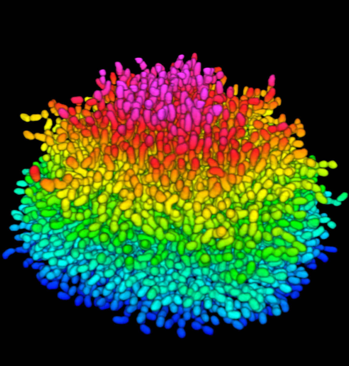
Previous efforts to delve into how the cells in a burgeoning biofilm interact had failed because of insufficient optical resolution; basically, what one cell was doing in the opaque mass could not be distinguished from its neighbors. 

The Princeton researchers overcame this problem in several ways. First, they genetically modified the bacterial strain so the cells produced proteins that glow brightly when illuminated by specific colors of light. The proteins selected offer the brightest available fluorescence, making each cell easier to pick out, while reducing the intensity of potentially cell-damaging light required for the experiment.

The team then used a confocal microscope, a device that focuses on a single portion of a specimen from a certain distance. By making hundreds of such observations, images can be stacked together to create a three-dimensional image of the entire specimen. “It’s like looking deep into the interior of a biofilm without having to slice it open,” said Yan.

Another boost for the research team came from computer algorithms originally developed for fields like materials science. The algorithms differentiated closely clustered sources of light, in this case the many bunched-up V. cholerae cells in a thickening biofilm. 

What the Princeton team saw was remarkable. At first, the bacterial colony expanded horizontally on the given surface in the experiment. As each cell split, the resulting daughter cells firmly attached to the surface alongside their parent cells. Squeezed by increasing numbers of offspring bacteria, however, the cells at the heart of expanding colony were forced to detach from the surface and point vertically. The bacterial colony thus went from a flat, two-dimensional mass to an expanding, three-dimensional blob, all held together by gunk in the developing biofilm.

The Princeton team dug a bit deeper into the genetics behind this cellular behavior. A single gene, dubbed RbmA, is key to behavior in which new cells connect in such a way to develop a three-dimensional biofilm. When the researchers deactivated the gene, a big, diffuse and floppy biofilm formed. When RbmA performed as normal, though, a denser, stronger biofilm resulted as the cells stayed linked to each other. Thus, RbmA provides the biofilm its resilience, providing insight into a potential Achilles heel that could be targeted for therapeutic intervention.

Ongoing work is now measuring the physical forces experienced by cells uplifting at the biofilm’s center so the overall mechanics can be precisely worked out. “We are currently trying to develop a mathematical model for how the bacterial colony grows in time and how the spatial features are linked to typical mechanical features of the biofilm,” said Stone. 

Disinfection tolerance of C. perfringens on farms and in processing

Clostridium perfringens is a Gram-positive, aerotolerant anaerobic spore-forming bacterium that causes a wide variety of diseases in humans and animals, primarily as a result of its ability to produce many different toxins (Markey et al., 2013). In humans, C. perfringens is responsible for gas gangrene, enteritis necroticans, food poisoning, and antibiotic-associated diarrheas ( Myers et al., 2006). Currently, C. perfringens type A food poisoning ranks as the second most commonly reported foodborne illness in Canada (Thomas et al., 2013).

c-perfringens-farmIn poultry, avian-specific C. perfringens strains cause necrotic enteritis, an economically significant poultry disease that costs the global industry over $2 billion annually in losses and control measures (Stanley et al., 2014). In some countries, this disease appears to be on the rise because of removal of antibiotic growth promoters (Stanley et al., 2014). C. perfringens is also a cause of various enterotoxemia in other animal species. Isolates of animal origin constitute a risk for transmission to humans through the food chain.

In order to persist in the environment, many bacteria have evolved the ability to form biofilms (Davey and O’Toole, 2000 and Jefferson, 2004). In fact, the predominant organizational state of bacteria in nature is biofilms (Costerton, 1999). Important features of cells in biofilms include: aggregation in suspension or on solid surfaces, increased antibiotic tolerance, and resistance to physical and environmental stresses (Davey and O’Toole, 2000, Davies, 2003 and Hall-Stoodley and Stoodley, 2009). It is now generally accepted that the biofilm growth mode induces bacterial tolerance to disinfection that can lead to substantial economic and health concerns (Bridier et al., 2011). Although the precise mechanism of such tolerance remains unclear, a review has recently discussed the subject as a multifactorial process involving the spatial organization of the biofilm (Bridier et al., 2011). More recently, we, and others, have described the formation of biofilms in C. perfringens (Charlebois et al., 2014 and Varga et al., 2006). We demonstrated that the biofilm formed by C. perfringens could protect the cells from an exposure to atmospheric oxygen and to high concentrations of antibiotics and anticoccidial agents ( Charlebois et al., 2014). It has also been observed that the biofilm formed by C. perfringens could protect the cells from an exposure to 10 mM of hydrogen peroxide even though this bacterium is catalase-negative (Varga et al., 2006). The capacity of C. perfringens to be part of dual- or multi-species biofilm has recently been reviewed ( Pantaleon et al., 2014) and C. perfringens biofilm was detected in many types of multi-species biofilm including biliary stents (Leung et al., 2000 and Pantaleon et al., 2014).

However, susceptibilities of C. perfringens mono- and dual-species biofilms exposed to most disinfectants are currently unknown. This study was undertaken to investigate the tolerance of C. perfringens mono- and dual-species biofilms to disinfectants used in farms and food processing environments.

Tolerance of Clostridium perfringens biofilms to disinfectants commonly used in the food industry

Journal of Food Microbiology

Volume 62, April 2017, p. 32-38

Charlebois, Audrey. Et al.

http://www.sciencedirect.com/science/article/pii/S0740002015300927

Listeria can be sticky

This study evaluated the occurrence of L. monocytogenes in the processing environment of a butcher shop, and the in vitro adhesion capacity and sensitivity of isolates to two sanitizers: A (Mister MaxDG1, chlorine based) and B (B-Quart Sept, quaternary ammonium based).

rolling_stones-sticky_fingersOf the total of 40 samples, 75% were positive for Listeria spp. and 22.5% for L. monocytogenes. 20 isolates were from serogroup 1/2c or 3c, with positive results for all tested virulence genes. All isolates presented adhesion potential. The evaluated sanitizers had the potential to inhibit isolates growth and adhesion, and removed formed biofilms. After evaluation, the sanitizers were adopted by the butcher shop in its sanitation routine, being effective against L. monocytogenes.

Collected data allowed identification of adhesion potential by L. monocytogenes and the effectiveness of the tested sanitizers to control contamination by this pathogen.

Listeria spp. contamination in a butcher shop environment and Listeria monocytogenes adhesion ability and sensitivity to food-contact surface sanitizers

Journal of Food Safety, DOI: 10.1111/jfs.12313, ahead of print

DAL Silva, AC Camargo, SD Todorov, LA Nero

http://onlinelibrary.wiley.com/doi/10.1111/jfs.12313/abstract;jsessionid=302167477C8A0B64C7E52E6E08696398.f03t02

STEC E. coli and biofilm production

The objectives of this study were to characterize the phenotype and genotype of 36 non-O157 Shiga toxin–producing Escherichia coli (STEC) strains isolated from humans, ovines, or bovines, including the top 6 (O26, O45, O103, O111, O121, and O145) and three other serogroups implicated in serious illness (O91, O113, and O128).

e,coli.biofilmBiofilms were formed by all strains with intermediate to strong biofilm producers (n = 24) more common at 22°C than at 37°C (p < 0.001) and 48 and 72 h (p < 0.001) than 24 h of incubation time. Biofilm-forming potential differed by serogroup and origin with O113 and human strains exhibiting the highest potential (p < 0.001). Biofilm-associated genes, csgA/csgD/crl/fimH (100%), flu (94%), rpoS (92%), ehaAα (89%), and cah (72%), were most prevalent, while mlrA (22%) and ehaAβ (14%) were least prevalent, although there was no clear compliment of genes associated with strains exhibiting the greatest biofilm-forming capacity.

Among 12 virulence genes screened, iha and ehxA were present in 92% of the strains. The occurrence of stx1 in the top 6 serogroups (8/12, 67%) did not differ (p = 0.8) from other serogroups (17/24, 71%), but stx2 was less likely (confidence interval [CI] = 0.14–1.12; p = 0.04) to be in the former (9/24, 38%) than the latter (9/12, 75%). Excluding serogroups, O91 and O121, at least one strain per serogroup was resistant to between three and six antimicrobials. Streptomycin (31%), sulfisoxazole (31%), and tetracycline (25%) resistance was most common and was 35–50% less likely (p < 0.05) in human than animal strains.

All non-O157 STEC strains were able to form biofilms on an abiotic surface, with some exhibiting resistance to multiple antimicrobials. Potential as a reservoir of antimicrobial resistance genes may be another hazard of biofilms in food-processing plants. As a result, future strategies to control these pathogens may include measures to prevent biofilms.

Biofilm formation, virulence gene profiles, and antimicrobial resistance of nine serogroups of non-O157 shiga toxin-producing E. coli

Wang, K. Stanford, T.A. McAllister, R.P. Johnson, J. Chen, H. Hou, G. Zhang, and Y.D. Niu

Foodborne Pathogens and Disease, Volume 13, Number 6, March 2016, Pages 1-9, DOI: 10.1089/fpd.2015.2099

http://online.liebertpub.com/doi/abs/10.1089/fpd.2015.2099